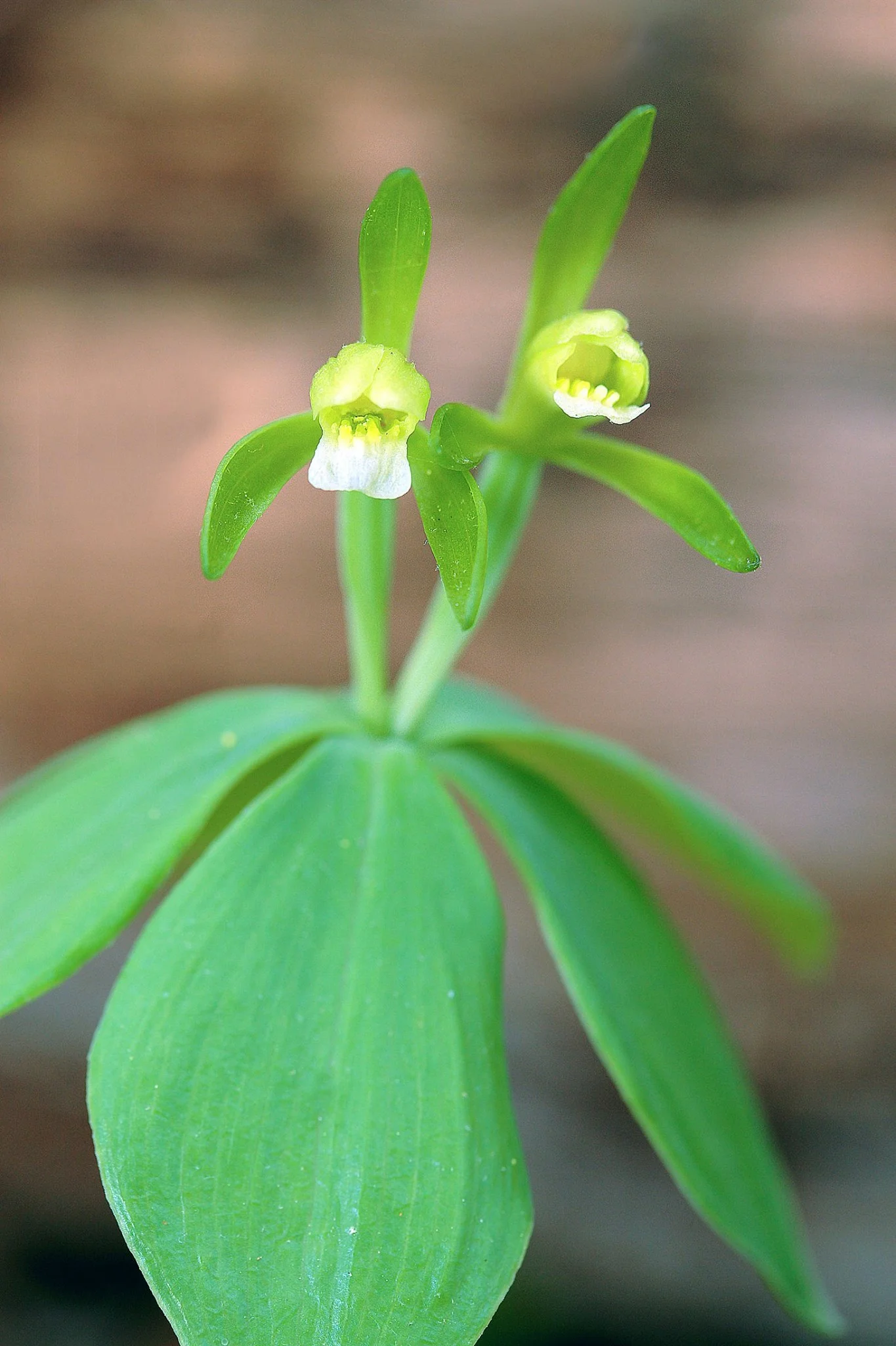
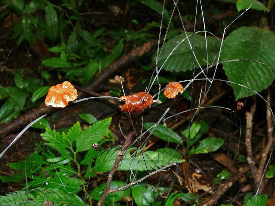
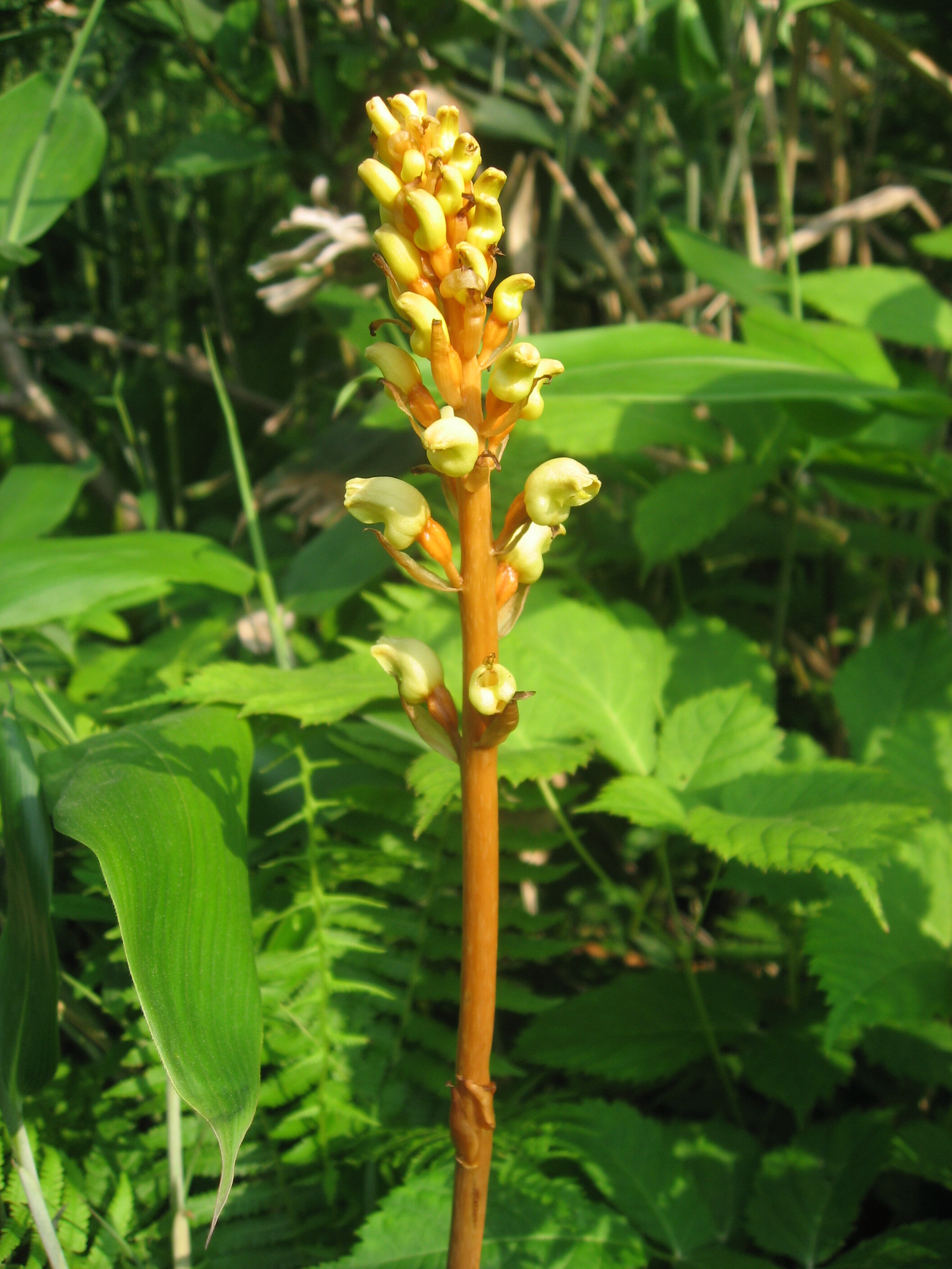
A Newly Described Fungus That Mimics Flowers

(A) Young yellow-orange pseudoflower. (B) Mature pseudoflower. (C) Longitudinal section of an infected X. surinamensis inflorescence. (D) Healthy yellow flower of X. surinamensis shown for comparison. [SOURCE]

(A) Young yellow-orange pseudoflower. (B) Mature pseudoflower. (C) Longitudinal section of an infected X. surinamensis inflorescence. (D) Healthy yellow flower of X. surinamensis shown for comparison. [SOURCE]

Imagine there was a fungus that was able to hijack human reproductive structures so that it could reproduce. Though this sounds like the basis of a strange science fiction story, a similar situation to this has just been described from Guyana between two species of yellow-eyed grass (Xyris setigera & X. surinamensis) and a newly described species of fungus called Fusarium xyrophilum.

Fungi that hijack plant reproductive systems are pretty rare in nature, especially when you consider the breadth of interactions between these two branches on the tree of life. What makes this newly described case of floral hijacking so remarkable is the complexity of the whole process. It all begins when an infected Xyris host begins to produce its characteristically lanky inflorescence.

At first glance, nothing would appear abnormal. The floral spike elongates and the inflorescence at the tip gradually matures until the flowers are ready to open. Even when the “flower” begins to emerge from between the tightly packed bracts the process seems pretty par for the course. Gradually a bright yellow, flower-like structure bursts forth, looking very much like how a bright yellow Xyris flower should look. However, a closer inspection of an infected plant would reveal something very different indeed.

Instead of petals, anthers, and a pistil, infected inflorescences produce what is called a pseudoflower complete with petal-like structures. This pseudoflower is not botanical at all. It is made entirely by the Fusarium fungus. Amazingly, these similarities are far from superficial. When researchers analyzed these pseudoflowers, they found that they are extremely close mimics of an actual Xyris flower in more than just looks. For starters, they produce pigments that reflect UV light in much the same way that actual flowers do. They also emit a complex suite of volatile scent compounds that are known to attract pollinating insects. In fact, at least one of those compounds was an exact match to a scent compound produced by the flowers of these two Xyris species.

So, why would a fungus go through all the trouble of mimicking its hosts flowers so accurately? For sex, of course! This species of Fusarium cannot exist without its Xyris hosts. However, Xyris don’t live forever and for the cycle to continue, Fusarium must go on to infect other Xyris individuals. This is where those pseudoflowers come in. Because they so closely match actual Xyris flowers in both appearance and smell, pollinating bees treat them just like flowers. The bees land on and investigate the fungal structure until they figure out there is no reward. No matter, they have already been covered in Fusarium spores.

As the bees visit other Xyris plants in the area, they inevitably deposit spores onto each plant they land on. Essentially, they are being coopted by the fungus in order to find new hosts. By mimicking flowers, the Fusarium is able to hijack plant-pollinator interactions for its own reproduction. It is not entirely certain at this point just how specific this fungus is to these two Xyris species. A search for other potential hosts turned up only a single case of it infecting another Xyris. It is also uncertain as to how much of an impact this fungus has on Xyris reproduction. Though the fungus effectively sterilizes its host, researchers did make a point to mention that Xyris populations may actually benefit from having a few infected plants as the pseudoflowers last much longer than the actual flowers and therefore could serve to attract more pollinators to the area over time. Who knows what further investigations into the ecology of this bizarre system will reveal.

Photo Credit: [1]

Further Reading: [1]

How radioactive carbon from nuclear bomb tests can tell us what parasitic orchids are eating

Yoania japonica. Photo by Qwert1234 licensed by CC BY-NC-SA 4.0

Yoania japonica. Photo by Qwert1234 licensed by CC BY-NC-SA 4.0

Historically, non-photosynthetic plants were defined as saprotrophs. It was thought that, like fungi, such plants lived directly off of decaying materials. Advances in our understanding have since revealed that parasitic plants don’t do any of the decaying themselves. Instead, those that aren’t direct parasites on the stems and roots of other plants utilize a fungal intermediary. We call these plants mycoheterotrophs (fungus-eaters). Despite recognition of this strangely fascinating relationship, we still have much to learn about what kinds of fungi these plants parasitize and where most of the nutritional demands are coming from.

It is largely assumed that most mycoheterotrophic plants are parasitic on mycorrhizal fungi. This would make them indirect parasites on other photosynthetic plants. The mycorrhizal fungi partner with photosynthetic plants, exchanging soil nutrients for carbon made by the plant during photosynthesis. However, this is largely assumed rather than tested. New research out of Japan has shown a light on what is going on with some of these parasitic relationships and the results are a bit surprising. What’s more, the methods they used to better understand these parasitic relationships are pretty clever to say the least.

Cyrtosia septentrionalis Photo by Qwert1234 licensed by CC BY-NC-SA 4.0

Cyrtosia septentrionalis Photo by Qwert1234 licensed by CC BY-NC-SA 4.0

Photosynthesis involves the uptake of and subsequent breakdown of CO2 from the atmosphere. The carbon from CO2 is then used to build carbohydrates, which form the backbone of most plant tissues. Not all carbon is created equal, however, and by looking at ratios of different carbon isotopes in living tissues, scientists can better understand where the carbon came from. For this research, scientists utilized an isotope of carbon called 14C.

Eulophia zollingeri photo by Vinayraja licensed by CC BY-NC-SA 3.0

Eulophia zollingeri photo by Vinayraja licensed by CC BY-NC-SA 3.0

14C is special because it is not as common in our atmosphere as other isotopes of carbon such as 12C and 13C. One of the biggest sources of 14C in our atmosphere were nuclear bomb explosions. From the 1950’s until the Partial Nuclear Test Ban in 1963, atomic bomb tests were a regular occurrence. During that time period, the concentration of 14C in our atmosphere greatly increased. Any organism that was fixing carbon into its tissues during that span of time will contain elevated levels of 14C compared to the other carbon isotopes. Alternatively, anything fixing carbon today, say via photosynthesis, will have considerably reduced levels of 14C in its tissues.

Gastrodia elata Photo by Qwert1234 licensed by CC BY-NC-SA 4.0

Gastrodia elata Photo by Qwert1234 licensed by CC BY-NC-SA 4.0

By looking at the ratios of 14C in the tissues of parasitic plants, scientists reasoned that they could assess the age of the carbon being utilized. If more 14C was present, the source of the carbon could not come from today’s atmosphere and therefore not from recent photosynthesis. Instead, it would have to come from older sources like decaying wood of long-dead trees. In other words, if parasitic plants were high in 14C, then the scientists could reasonably conclude that they were parsitizing wood-decaying saprotrophic fungi. If the plants were high in 12C or 13C, then they could conclude that they were partnering with mycorrhizal fungi instead, which were obtaining carbon from present-day photsynthesis.

The researchers looked at 10 different species of parasitic plants across Japan, most of which were orchids. They analyzed their tissues and ran analyses on the carbon molecules within. What they found is that 6 out of the 10 plants contained much higher levels of 12C and 13C in their tissues, which points to mycorrhizal fungi as their host. However, for the 4 remaining species (Gastrodia elata, Cyrtosia septentrionalis, Yoania japonica and Eulophia zollingeri), the ratios of 14C were considerably higher, meaning their host fungi were eating dead wood, not partnering with photosynthetic plants near by.

Indeed, it appears that at least some mycoheterotrophic plants are benefiting from saprotrophic rather than mycorrhizal fungi. Those early assumptions into the livelihood of such plants were not as far off the mark after all. This is very exciting research that opens the door to a much deeper understanding of some of the strangest plants on our planet.

LEARN MORE ABOUT MYCOHETEROTROPHIC PLANTS IN EPISODE 234 OF THE IN DEFENSE OF PLANTS PODCAST

Photo Credits: [1] [2] [3] [4] [5]

Further Reading: [1] [2]

The Fungus-Mimicking Mouse Plant

18156089_1751541684872689_3055288262507833768_o.jpg

The mouse plant (Arisarum proboscideum) is, to me, one of the most charming aroids in existence. Its small stature and unique inflorescence are a joy to observe. It is no wonder that this species has attained a level of popularity among those of us who enjoy growing oddball plants. Its unique appearance may be reason enough to appreciate this little aroid but its pollination strategy is sure to seal the deal.

The mouse plant is native to shaded woodlands in parts of Italy and Spain. It is a spring bloomer, hitting peak flowering around April. It has earned the name “mouse plant” thanks to the long, tail-like appendage that forms at the end of the spathe. That “tail” is the only part of the inflorescence that sticks up above the arrow-shaped leaves. The rest of the structure is presented down near ground level. From its stature and position, to its color, texture, and even smell, everything about the inflorescence is geared around fungal mimicry.

The mouse plant is pollinated by fungus gnats. However, it doesn’t offer them any rewards. Instead, it has evolved a deceptive pollination syndrome that takes advantage of a need that all living things strive to attain - reproduction. To draw fungus gnats in, the mouse plant inflorescence produces compounds that are said to smell like fungi. Lured by the scent, the insects utilize the tail-like projection of the spathe as a sort of highway that leads them to the source.

Once the fungus gnats locate the inflorescence, they are presented with something incredibly mushroom-like in color and appearance. The only opening in the protective spathe surrounding the spadix and flowers is a tiny, dark hole that opens downward towards the ground. This is akin to what a fungus-loving insect would come to expect from a tiny mushroom cap. Upon entering, the fungus gnats are greeted with the tip of the spadix, which has come to resemble the texture and microclimate of the underside of a mushroom.

Anatomy of a mouse plant inflorescence [SOURCE]

Anatomy of a mouse plant inflorescence [SOURCE]

This is exactly what the fungus gnats are looking for. After a round of courtship and mating, the fungus gnats set to work laying eggs on the tip of the spadix. Apparently the tactile cues are so similar to that of a mushroom that the fungus gnats simply don’t realize that they are falling victim to a ruse. Upon hatching, the fungus gnat larvae will not be greeted with a mushroomy meal. Instead, they will starve and die within the wilting inflorescence. The job of the adult fungus gnats is not over at this point. To achieve pollination, the plant must trick them into contacting the flowers themselves.

Both male and female flowers are located down at the base of the structure. As you can see in the pictures, the inflorescence is two-toned - dark brown on top and translucent white on the bottom. The flowers just so happen to sit nicely within the part of the spathe that is white in coloration. In making a bid to escape post-mating, the fungus gnats crawl/fly towards the light. However, because the opening in the spathe points downward, the lighted portion of the structure is down at the bottom with the flowers.

The leaves are the best way to locate these plants. Photo by Meneerke bloem licensed under CC BY-SA 4.0

The leaves are the best way to locate these plants. Photo by Meneerke bloem licensed under CC BY-SA 4.0

Confused by this, the fungus gnats dive deeper into the inflorescence and that is when they come into contact with the flowers. Male and female flowers of the mouse plants mature at the exact same time. That way, if visiting fungus gnats happen to be carrying pollen from a previous encounter, they will deposit it on the female flowers and pick up pollen from the male flowers all at once. It has been noted that very few fungus gnats have ever been observed within the flower at any given time so it stands to reason that with a little extra effort, they are able to escape and with any luck (for the plant at least) will repeat the process again with neighboring individuals.

The mouse plant does not appear to be self-fertile so only pollen from unrelated individuals will successfully pollinate the female flowers. This can be a bit of an issue thanks to the fact that plants also reproduce vegetatively. Large mouse plant populations are often made up of clones of a single individual. This may be why rates of sexual reproduction in the wild are often as low as 10 - 20%. Still, it must work some of the time otherwise how would such a sophisticated form of pollination syndrome evolve in the first place.

Photo Credit: [1] [2] [3]

Further Reading: [1] [2]

How a Giant Parasitic Orchid Makes a Living

Photo by mutolisp licensed under CC BY-NC-SA 2.0

Photo by mutolisp licensed under CC BY-NC-SA 2.0

Imagine a giant vine with no leaves and no chlorophyll scrambling over decaying wood and branches of a warm tropical forest. As remarkable as that may seem, that is exactly what Erythrorchis altissima is. With stems that can grow to upwards of 10 meters in length, this bizarre orchid from tropical Asia is the largest mycoheterotrophic plant known to science.

Mycoheterotrophs are plants that obtain all of their energy needs by parasitizing fungi. As you can probably imagine, this is an extremely indirect way for a plant to make a living. In most instances, this means the parasitic plants are stealing nutrients from the fungi that were obtained via a partnership with photosynthetic plants in the area. In other words, mycoheterotrophic plants are indirectly stealing from photosynthetic plants.

In the case of E. altissima, this begs the question of where does all of the carbon needed to build a surprising amount of plant come from? Is it parasitizing the mycorrhizal network associated with its photosynthetic neighbors or is it up to something else? These are exactly the sorts of questions a team from Saga University in Japan wanted to answer.

Photo by mutolisp licensed under CC BY-NC-SA 2.0

Photo by mutolisp licensed under CC BY-NC-SA 2.0

All orchids require fungal partners for germination and survival. That is one of the main reasons why orchids can be so finicky about where they will grow. Without the fungi, especially in the early years of growth, you simply don't have orchids. The first step in figuring out how this massive parasitic orchid makes its living was to identify what types of fungi it partners with. To do this, the team took root samples and isolated the fungi living within.

By looking at their DNA, the team was able to identify 37 unique fungal taxa associated with this species. Most surprising was that a majority of those fungi were not considered mycorrhizal (though at least one mycorrhizal species was identified). Instead, the vast majority of the fungi associated with with this orchid are involved in wood decay.

Stems climbing on fallen dead wood (a) or on standing living trees (b). A thick and densely branched root clump (c) and thin and elongate roots (d) [Source]

Stems climbing on fallen dead wood (a) or on standing living trees (b). A thick and densely branched root clump (c) and thin and elongate roots (d) [Source]

To ensure that these wood decay fungi weren't simply partnering with adult plants, the team decided to test whether or not the wood decay fungi were able to induce germination of E. altissima seeds. In vitro germination trials revealed that not only do these fungi induce seed germination in this orchid, they also fuel the early growth stages of the plant. Further tests also revealed that all of the carbon and nitrogen needs of E. altissima are met by these wood decay fungi.

These results are amazing. It shows that the largest mycoheterotrophic plant we know of lives entirely off of a generalized group of fungi responsible for the breakdown of wood. By parasitizing these fungi, the orchid has gained access to one of the largest pools of carbon (and other nutrients) without having to give anything back in return. It is no wonder then that this orchid is able to reach such epic proportions without having to do any photosynthesizing of its own. What an incredible world we live in!

Photo by mutolisp licensed under CC BY-NC-SA 2.0

Photo by mutolisp licensed under CC BY-NC-SA 2.0

Photo Credits: [1] [2]

Further Reading: [1]

How Trees Fight Disease

27329418_10101829159766245_2047871682_o.jpg

Plants do not have immune systems like animals. Instead, they have evolved an entirely different way of dealing with infections. In trees, this process is known as the "compartmentalization of decay in trees" or "CODIT." CODIT is a fascinating process and many of us will recognize its physical manifestations.

In order to understand CODIT, one must know a little something about how trees grow. Trees have an amazing ability to generate new cells. However, they do not have the ability to repair damage. Instead, trees respond to disease and injury  by walling it off from their living tissues. This involves three distinct processes. The first of these has to do with minimizing the spread of damage. Trees accomplish this by strengthening the walls between cells. Essentially this begins the process of isolating whatever may be harming the living tissues.

This is done via chemical means. In the living sapwood, it is the result of changes in chemical environment within each cell. In heartwood, enzymatic changes work on the structure of the already deceased cells. Though the process is still poorly understood, these chemical changes are surprisingly similar to the process of tanning leather. Compounds like tannic and gallic acids are created, which protect tissues from further decay. They also result in a discoloration of the surrounding wood. 

The second step in the CODIT process involves the construction of new walls around the damaged area. This is where the real compartmentalization process begins. The cambium layer changes the types of cells it produces around the area so that it blocks that compartment off from the surrounding vascular tissues. These new cells also exhibit highly altered metabolisms so that they begin to produce even more compounds that help resist and hopefully stave off the spread of whatever microbes may be causing the injury. Many of the defects we see in wood products are the result of these changes.

CODIT.JPG

The third response the tree undergoes is to keep growing. New tissues grow around the infected compartment and, if the tree is healthy enough, will outpace further infection. You see, whether its bacteria, fungi, or a virus, microbes need living tissues to survive. By walling off the affected area and pumping it full of compounds that kill living tissues, the tree essentially cuts off the food supply to the disease-causing organism. Only if the tree is weakened will the infection outpace its ability to cope.

Of course, CODIT is not 100% effective. Many a tree falls victim to disease. If a tree is not killed outright, it can face years or even decades of repeated infection. This is why we see wounds on trees like perennial cankers. Even if the tree is able to successfully fight these repeat infections over a series of years, the buildup of scar tissues can effectively girdle the tree if they are severe enough.

CODIT is a well appreciated phenomenon. It has set the foundation for better tree management, especially as it relates to pruning. It is even helping us develop better controls against deadly invasive pathogens. Still, many of the underlying processes involved in this response are poorly understood. This is an area begging for deeper understanding.

Photo Credits: kaydubsthehikingscientist & Alex Shigo

Further Reading: [1]

The White Walnut

Photo by Dan Mullen licensed under CC BY-NC-ND 2.0

Photo by Dan Mullen licensed under CC BY-NC-ND 2.0

I must admit, I am not very savvy when it comes to trees. I love and appreciate them all the same, however, my attention is often paid to the species growing beneath their canopy. last summer changed a lot of that. I was very lucky to be surrounded by people that know trees quite well. Needless to say I picked up a lot of great skills from them. Despite all of this new information knocking around in my brain, there was one tree that seemed to stand out from the rest and that species is Juglans cinerea.

Afternoons and evenings at the research station were a time for sharing. We would all come out of the field each day tired but excited. The days finds were recounted to eager ears. Often these stories segued into our goals for the coming days. That is how I first heard of the elusive "white walnut." I had to admit, it sounded made up. Its as if I was being told a folktale of a tree that lived in the imagination of anyone who spent too much time in the forest. 

Only a handful of people knew what it was. I listened intently for a bit, hoping to pick up some sort of clue as to what exactly this tree was. Finally I couldn't take it any longer so I chimed in and asked. As it turns out, the white walnut is a tree I was already familiar with, though not personally. Another common name for this mysterious tree is the butternut. Ah, common names. 

I instantly recalled a memory from a few years back. A friend of mine was quite excited about finding a handful of these trees. He was very hesitant to reveal the location but as proof of his discovery he produced a handful of nuts that sort of resembled those of a black walnut. These nuts were more egg shaped and not nearly as large. Refocusing on the conversation at hand, I now had a new set of questions. Why was this tree so special? Moreover, why was it so hard to find?

The white walnut has quite a large distribution in relation to all the excitement. Preferring to grow along stream banks in well-drained soils, this tree is native from New Brunswick to northern Arkansas. Its leaflets are downy, its bark is light gray to almost silver, and it has a band of fuzzy hairs along the upper margins of the leaf scars. Its a stunning tree to say the least. 

Sadly, it is a species in decline. As it turns out, the excitement surrounding this tree is due to the fact that finding large, robust adults has become a somewhat rare occurrence. Yet another casualty of the global movement of species from continent to continent, the white walnut is falling victim to an invasive species of fungus known scientifically as Sirococcus clavigignenti-juglandacearum

The fungus enters the tree through wounds in the bark and, through a complex life cycle, causes cankers to form. These cankers open the tree up to subsequent infections and eventually girdle it. The fungus was first discovered in Wisconsin but has now spread throughout the entire range of the tree. The losses in Wisconsin alone are staggering with an estimated 90% infection rate. Farther south in the white walnuts range, it is even worse. Some believe it is only a matter of time before white walnut becomes functionally extinct in areas such as the Carolinas. No one knows for sure where this fungus came from but Asia is a likely candidate.

A sad and all too common story to say the least. It was starting to look like I was not going to get a chance to meet this tree in person... ever. My luck changed a few weeks later. My friend Mark took us on a walk near a creek and forced us to keep our eyes on the canopy. We walked under a tree and he made sure to point out some compound leaves. With sunlight pouring through the canopy we were able to make out a set of leaves with a subtle haze around the leaf margins. We followed the leaves to the branches and down to the trunk. It was silvery. There we were standing under a large, healthy white walnut. The next day we stumbled across a few young saplings in some of our vegetation plots. All is not lost. I can't speak for the future of this species but I feel very lucky to have seen some healthy individuals. With a little bit of luck there may be hope of resistance to this deadly fungus. Only time will tell. 

Photo Credit: Dan Mullen (http://bit.ly/2br2F0Z)

Further Reading:
http://bit.ly/2b8GiMV

http://bit.ly/2aLUdMD

Cedar-Apple Rust

Photo by Rocky Houghtby licensed under CC BY 2.0

Photo by Rocky Houghtby licensed under CC BY 2.0

I have had my eye on these strange brown golf ball shaped growths growing on the twigs of a cedar in my neighborhood for about a year now. I first took notice of them late last spring. They looked pretty nasty but I knew they had to be something interesting. Indeed, “interesting” doesn't even come close to describing their true nature. 

These odd little growths are actually a single stage in the complex life cycle of a group of fungi in the genus Gymnosporangium. Collectively they are referred to as cedar-apple galls. Its a group of fungi whose hosts include junipers and relatives of the apple. Wherever these two lineages coexist you are bound to find this fungus. 

Gymnosporangium exhibit a fascinating life cycle that includes multiple hosts. The golf ball shaped galls will appear on the twigs of a juniper nearly a year after being infected with spores. They grow in size until they reach a point in which they will barely fit in the palm of your hand, though not all reach such proportions. The gall itself is covered in a series of uniform depressions, making it look a little out of place in a natural setting. After a year on a juniper tree, the galls enter into their next stage of development. 

Photo by klm185 licensed under CC BY 2.0

Photo by klm185 licensed under CC BY 2.0

Usually triggered by the first warm rains of spring, strange gelatinous protrusions start to poke out of each depression on the gall's surface. These protrusions continue to swell until the entire gall is covered in bright orange, finger-like masses. These are where the spores are produced. The spores, however, cannot infect another juniper. Instead, they need to land on the next host to complete their life cycle. 

If the spores land on a member of the family Rosaceae (species within the genus Malus are preferred), then the second stage of the life cycle begins. Spores can germinate on both the leaves and the fruit but instead of turning into a large brown gall, they take on a different appearance. This is what makes this fungus readily apparent as a type of rust. A patch of orange will begin to grow. Upon closer inspection one can see that the orange patch is actually a series of small cup-like structures full of spores. 

Come fall, the spores are ready to be dispersed by wind. With any luck, they will land back on a juniper tree and the cycle will start anew. Because of its propensity for apple crops, cedar-apple rust fungi are considered to be a serious pest in apple orchards. In a more natural setting, however, it is a bizarrely unique fungus worth looking for.

Photo Credits: [1] [2]

Further Reading: [1] [2]

On Fungi and Forest Diversity

One simply can't talk about plants without eventually talking about fungi. The fact of the matter is the vast majority of plant species rely on fungal interactions for survival. This mutualistic relationship is referred to as mycorrhizal. Fungi in the soil colonize the root system of plants and assist in the acquisition of nutrients such as nitrogen and phosphorus. In return, most photosynthetic plants pay their mycorrhizal symbionts with carbohydrates. 

There are two major categories of mycorrhizal fungi - ectomycorrhizae (EMF) and arbuscular mycorrhizae (AMF). Though there are a variety of different species of fungi that fall into either of these groups, their strategies are pretty much the same. EMF make up roughly 10% of all the known mycorrhizal symbionts. The prefix "ecto" hints at the fact that these fungi form on the outside of root cells. They form a sort of sheath that encases the outside of the root as well as a "hartig net" around the outside of individual cells within the root cortex. AMF, on the other hand, literally penetrate the root cells and form two different kinds of structures once inside. One of these structures looks like the crown of a tree, hence the term "arbuscular." What's more, they are considered the oldest mycorrhizal group to have evolved. 

The type of mycorrhizal fungi a plant partners with has greater implications that simple nutrient uptake. Evidence is now showing that the dominant fungi of a region can actually influence the overall health and diversity forest ecosystems. The mechanism behind this has a lot to do with the two different categories discussed above. 

Researchers have discovered that trees partnering with AMF experience negative feedbacks in biomass whereas those that partner with EMF experience positive feedbacks in biomass. When grown in soils that previously harbored similar tree species, trees that partnered with AMF grew poorly whereas trees that partnered with EMF grew much better. Additionally, by repeating the experiments with seedlings, researchers found that seedling survival was reduced for AMF trees whereas seedling survival increased in EMF trees. 

What is going on here? If mycorrhizae are symbionts, why would there be any detrimental effects? The answer to this may have something to do with soil pathogens. Thinking back to the major differences between EMF and AMF, you will remember that it comes down to the way in which they form their root associations. EMF form a protective sheath around the roots whereas AMF penetrate the cells.  As it turns out, this has major implications for pathogen resistance. Because they form a sheath around the entire root, EMF perform much better at keeping pathogens away from sensitive root tissues. The same can't be said for AMF. Researchers found that AMF trees experienced significantly more root damage when grown in soils that previously contained AMF trees. 

The differences in the type of feedback experienced by EMF and AMF trees can have serious consequences for tree diversity. Because EMF trees are healthier and experience increased seedling establishment in soils containing other EMF species, it stands to reason that this would lead to a dominance of EMF species, thus reducing the variety of species capable of establishing in that area. Conversely, areas dominated by AMF trees may actually be more diverse due to the reduction in fitness that would arise if AMF trees started to dominate. Though they are detrimental, the negative feedbacks experienced by AMF trees may lead to healthier and more diverse forests in the grand scheme of things. 

Infographic by [1]

Further Reading: [1]


Orchid Dormancy Mediated by Fungi

Photo by NC Orchid licensed under CC BY-NC 2.0

Photo by NC Orchid licensed under CC BY-NC 2.0

North America's terrestrial orchids seem to have mastered the disappearing act. When stressed, these plants can enter into a vegetative dormancy, existing entirely underground for years until the right conditions return for them to grow and bloom. Cryptic dormancy periods like this can make assessing populations quite difficult. Orchids that were happy and flowering one year can be gone the next... and the next... and the next...

How and why this dormancy is triggered has confused ecologists and botanists alike. Certainly stress is a factor but what else triggers the plant into going dormant? According to a recent paper published in the American Journal of Botany, the answer is fungal.

Orchids are the poster children for mycorrhizal symbioses. Every aspect of an orchid's life is dependent on these fungal interactions. Despite our knowledge of the importance of mycorrhizal presence in orchid biology, no one had looked at how the abundance of mycorrhizal fungi influenced the life history of these charismatic plants until now.

By observing the presence and abundance of a family of orchid associated fungi known as Russulaceae, researchers found that the abundance of mycorrhizal fungi in the environment is directly related to whether or not an orchid will emerge. The team focused on a species of orchid known commonly as the small whorled pogonia (Isotria medeoloides). Populations of this federally threatened orchid are quite variable and assessing their numbers is difficult.

The team found that the abundance of mycorrhizal fungi is not only related to prior emergence of these plants but could also be used as a predictor of future emergence. This has major implications for orchid conservation overall. It's not enough to simply protect orchids, we must also protect the fungal communities they associate with.

Research like this highlights the need for a holistic habitat approach to conservation issues. So many species are partners in symbiotic relationships and we simply can't value one partner over the other. If conditions change to the point that they no longer favor the mycorrhizal partner, it stands to reason that it would only be a matter of years before the orchids disappeared for good.

Photo Credit: NC Orchid

Further Reading: [1]

The Cranefly Orchid (Tipularia discolor)

RSCN0432.JPG

Look closely or you might miss it. Heck, even with close inspection you still run the risk of overlooking it. At this time of year, finding a cranefly orchid (Tipularia discolor) can present a bit of a challenge. At other times of the year the task is a bit easier. If you can find one in bloom, however, you are rewarded with, a unique orchid experience.

For most of the year, the cranefly orchid exists as a single leaf, which is produced in the fall and lasts until spring. It is thought that this orchid takes advantage of the dormancy of its neighbors by sucking up the light the canopy otherwise intercepts during the growing season. Any of you curious enough to look will have noticed that the underside of this leaf is deep purple in color. This very well may be an adaptation to take full advantage of light when it is available. There is some evidence that such coloration may help reflect light back up into the leaf, thus getting more out of what makes it to the forest floor. Evidence for this, however, is limited. It is far more likely that the purple coloration are pigments produced by the leaves that act as a sort of sun screen, shielding the sensitive photosynthetic machinery within from an overdose of sun as intense sun flecks dance across the forest floor.

By the end of spring, the single leaf has senesced. If energy stores were ample that year the plant will then flower. A lanky brown spike erupts from the ground. Its purple-green color is subtle yet beautiful. The flowers themselves are a bit odd, even by orchid standards. Whereas most orchid flowers exhibit satisfying bilateral symmetry, the flowers of the cranefly orchid are distinctly asymmetrical. The dorsal sepal, along with the lateral petals, are scrunched up on either side of the column. This has everything to do with its pollinators.

DSCN0414.JPG

The cranefly orchid has coopted nocturnal moths in the family Noctuidae for pollination. These moths find the flowers soon after they open and stick around only as long as there is nectar still present in the long nectar spurs. The asymmetry of the bloom causes the pollinia to attach to one of the moth's eyes. In this way the orchid is able to ensure that its pollen is not wasted on the blooms of other species.

As in all plants, the production of flowers is a costly business. Sexual reproduction is all about tradeoffs. It has been found that cranefly orchids that flowered and successfully produced fruit in one year were much less likely to do so in the next. What's more, the overall size of the plant (leaves and corms) were greatly reduced. Its hard to eek out an existence on the forest floor.

What I find most interesting about this species is where it tends to grow. Any old patch of ground simply won't do. Research indicates that the cranefly orchid requires rotting wood as a substrate. It's not so much the wood they require but rather the organisms that are decomposing it. Like all orchids, the cranefly cannot germinate and grow without mycorrhizal associations. They just happen to partner with fungi that also decompose wood. Such a relationship underscores the importance of decaying wood to forest health.

Further Reading: [1] [2] [3] [4]

A Fern With Flower Genes - An Odd Case of Horizontal Gene Transfer

Photo by Aaron Carlson licensed under CC BY-SA 2.0

Photo by Aaron Carlson licensed under CC BY-SA 2.0

When researchers at Harvard decided to take a look at the genome of the rattlesnake fern (Botrypus virginianum) they found something completely unexpected. Whereas one set of genes they looked at placed this species firmly in the family to which it belongs, Ophioglossaceae, two other genes placed it in the Loranthaceae, a completely unrelated family of flowering plants. What are flowering plant genes doing in a fern?

The rattlesnake fern is a ubiquitous species found throughout the northern hemisphere. It is believed to have evolved in Asia and then radiated outward using ancient land bridges that once connected the continents. At some point before this radiation occurred, the rattlesnake fern picked up some genes that were entirely foreign.

Horizontal gene transfer, the transfer of genes from one organism to another without reproduction, is nothing new in nature. Bacteria do it all the time. Even plants dabble in it every now and then. The surprising thing about this recently documented case is that it is the first discovery of horizontal gene transfer between an angiosperm and a fern. Up until this point, examples within the plant realm have been seen between ferns and hornworts as well as some parasitic plants and their hosts.

This is why the rattlesnake fern genome is so interesting. How did this occur? Though there is no way of telling for sure, researchers believe that one of two things could have happened. The first involves root parasitism. The family Loranthaceae is home to the mistletoes, a group of plants most famous for their parasitic nature. Although the majority of mistletoe species are stem parasites, at least three genera utilize root parasitism. It could be that an ancient species of mistletoe transferred some genes while parasitizing a rattlesnake fern.

This scenario seems to be the least likely of the two as no representatives of the root parasitic mistletoes currently exist in Asia, though it is entirely possible that some did at one time. The other possibility doesn't involve parasitism at all but rather fungi. Rattlesnake ferns are obligate mycotrophs and thus cannot live without certain species of mycorrhizal fungi. Perhaps the transfer of genes was achieved indirectly via a shared mycorrhizal network. This hypothesis is especially tantalizing because if it is found to be true, it would help explain many other examples of horizontal gene transfer that currently lack a mechanism. Only time and more research will tell.

Photo Credit: Aaron Carlson (http://bit.ly/1OAVhNZ)

Further Reading:
http://www.ncbi.nlm.nih.gov/pmc/articles/PMC1560187/

Rusty Mustards


image.jpg

Believe it or not, what you are seeing here is the same species of plant. The one on the left is the normal reproductive state of a Boechera (Arabis) mustard while the one on the right is the same species of mustard that has been infected by a rust fungus known as Puccinia monoica.

The interaction of these two species is interesting on so many levels. I spent an entire summer, along with my botanical colleagues, completely stumped as to what this strange orange-colored plant could be only to eventually find out that it was a mustard that has been hijacked! The fungus in question, P. monoica, is part of a large complex of interrelated rust fungi who are quite fond of mustards. The reason for this all boils down to reproduction.

The lifecycle of P. monoica begins when spores land on a young mustard plant and invade the host tissue. As they grow, they gain more and more nutrients from the mustard. Eventually the fungi effectively sterilizes the mustard and causes it to begin forming what are referred to as "pseudoflowers." The pseudoflowers are basically leaves that have been mutated by the fungus to look and smell a lot like other plants blooming in early summer.

The pseudoflowers produce a sticky, nectar-like substance that is very attractive to pollinators. The mimicry even goes as far as to produce yellowish pigments that reflect UV light, making them an even more irrisistable target for passing insects. On each pseudoflower are hundreds of small cups known as spermatogonia. These house the sex cells of the fungus. Visiting insects get covered in these sex cells, which they will then transfers to other infected plants thus achieving sexual reproduction for the fungus.

Still with me?

At this point, the pseudoflowers stop producing color and nectar and instead, the fused sex cells germinate into hyphae that begin to form specialized structures called "aecia." The aceia house the spores that will be responsible for infecting their secondary host plants, which are grasses. Spores blow about on the wind and, with a little luck, a few will land on a blade of grass. The spores germinate and infect the grass. From there, structures called "uredia" are formed that go on to produce even more spores to infect even more grass. Eventually, structures called "telia" are formed on the grass and the cycle finally comes full circle. The telia produce the spores that will go on to infect the original mustard host plants.

Whew! To have stumbled across an evolutionary drama such as this serves as a reminder of just how much in nature goes largely unnoticed every day.  

Further Reading: [1] [2] [3] [4]

Amber Fossils of Grain

image.jpg

In what may be one of the most interesting fossil discoveries in recent years, scientists from Oregon State University have described the earliest fossil evidence of grasses. Encased in 100 million year old amber this ancient grass spikelet suggests grasses were already around in the early to mid Cretaceous period. This is some 20 to 30 million years earlier than previous estimates for grass evolution. If that isn't cool enough, the grass appears to have been infected by a fungus related to ergot (the darker portion at the top), showing that this parasitism may be as old as grasses themselves. 

We humans have a long history with ergot's fondness for grasses. It is best known for producing the chemical precursors of LSD (as well as many other useful drugs) and has been implicated in some major historical events throughout our short time on this planet. However, suggesting that dinosaurs were getting high off the stuff is pushing it. Ergot likely evolved its chemical cocktail to deter herbivores from eating the grasses that it parasitizes. It has a bitter taste and cattle are said to avoid grasses that have been infected by it. It is quite possible that dinosaurs probably did the same thing. 

Either way, this finding represents a major milestone in the understanding of one of the most important plant families on the planet. Following the mass extinction at the end of the Cretaceous, grasses quickly rose to dominate roughly 20% of global vegetation. This little piece of amber now suggests that dinosaurs and their neighbors likely had a role in shaping this plant family. 

Photo Credit: Oregon State University

Further Reading: [1]